
Figure 1.
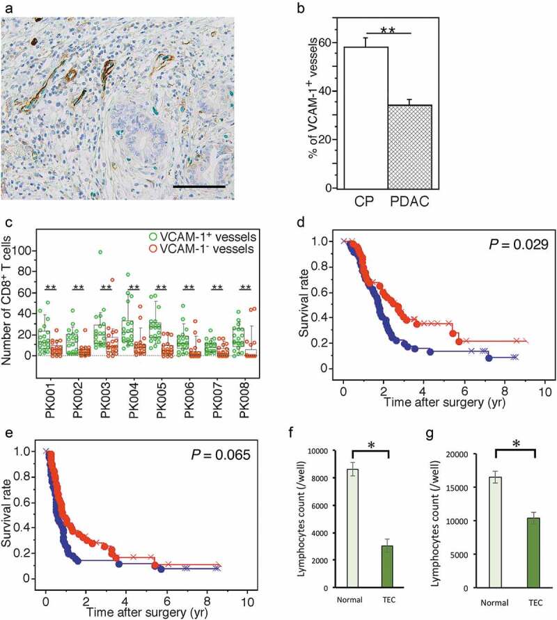
Endothelial cells in PDAC tissue are potentially dysfunctional for lymphocyte trafficking. (a) Double immunohistochemical detection of VCAM-1 (brown) and ERG1 (green) in PDAC tissue. High-power view. The bar indicates 100 µm. (b) Comparison of the percentage of vessels composed of VCAM-1+ endothelial cells among all vessels (%VCAM1+V) detected by evaluating ERG1+ endothelial cells in PDAC (n = 104) and CP tissue samples (n = 10). (c) Comparison of the numbers of tumor-infiltrating CD8+ T cells surrounding blood vessels composed of VCAM-1+ or VCAM-1− endothelial cells in PDAC tissue samples. Box plots and the scatter blots are combined. Whisker heights extend to 1.5 times the height of the box or, if no case/row has a value in that range, to the minimum and maximum values. The horizontal lines within the box indicate the median values. (d, e) Kaplan-Meier survival curves showing the comparison of OS (d) or DFS (e) between the high (red, n = 52) and low (blue, n = 52) %VCAM1+V groups of PDAC patients (n = 104). P-values were obtained from log-rank tests. “x” and “+” represent censoring and failure, respectively. (f, g) Adhesion assay evaluating the interaction between lymphocytes and TECs (f). Transmigration assay for lymphocytes migrating through an endothelial cell layer (g). Data are represented as the mean ± SD. Differences were analyzed by using the Mann-Whitney U test. *, P < .05 and **, P < .01