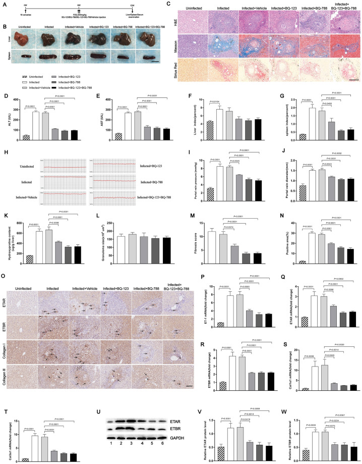
Fig 5. Endothelin receptor antagonists mediated reduction of hepatocellular damage.
Mice were infected percutaneously with 16 S. japonicum cercariae or remained uninfected. At 6 weeks post-infection, all infected mice were treated with praziquantel to kill the parasites and then received either endothelin receptor antagonists or vehicle and were necropsied at 12 weeks post-infection. (A) Time schedule for parasite infection and administration of anti-parasite drug or endothelin receptor antagonists and sample withdrawal. (B) Macrograph of livers and spleens from uninfected mice, infected mice and infected mice treated with endothelin receptor antagonists. Scale bar, 1 cm. (C) H&E, Masson’s trichrome and Sirius Red staining of liver sections. Scale bar, 200 μm. (D-E) Serum ALT and AST levels were measured (n = 5–7). (F-G) Liver and spleen indexes were determined (n = 7). (H) Measurement of hepatic portal venous pressure in vivo by RM6240BD. (I) Statistical analysis of hepatic portal vein pressure (n = 7). (J) Analysis of the portal vein diameter in vivo (n = 7). (K) Collagen content in livers determined as hydroxyproline content (n = 6). (L) Granuloma size measured based on H&E staining of liver sections (n = 7). (M) Fibrosis scores measured based on Masson’s trichrome staining of liver sections (n = 7). (N) The positive staining areas for Sirius Red were measured using IPP software (n = 7). (O) Representative immunohistochemical staining for ETAR, ETBR, collagen I and collagen III in infected livers. Black arrows indicate the ETRs, collagen I and collagen III positive cells. Scale bar, 100 μm. (P-T) qPCR analysis of the expression levels of ET-1, ETAR, ETBR, Col1α1 and Col3α1 in liver samples (n = 6). (U-W) ETAR and ETBR proteins were determined by western blotting. 1, uninfected; 2, infected; 3, infected + vehicle; 4, infected + BQ-123; 5, infected + BQ-788; 6, infected + BQ-123 + BQ-788. Image density was quantified using Image J analysis and normalized to GAPDH (n = 5). Data are represented as mean ± SEM of three independent experiments. Multiple comparisons were performed by one-way ANOVA with Tukey’s correction for comparison between two groups (D-G, I-N, P-T, V-W). Abbreviation: ALT: alanine aminotransferase; AST: aspartate aminotransferase.

