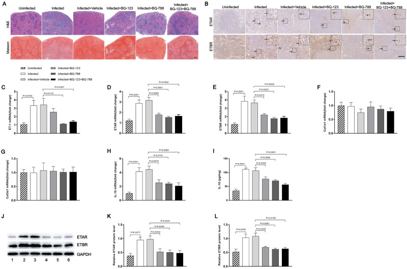
Fig 6. Endothelin receptor antagonists attenuated schistosomiasis-induced splenic fibrosis in mice.
(A) H&E and Masson’s trichrome staining of spleen sections. Scale bar, 200 μm. (B) Representative immunohistochemical staining for ETAR and ETBR in infected spleens. Insets show a higher magnification of the outlined area. Black arrows indicate the ETRs positive cells. Scale bar, 100 μm. (C-H) The expression levels of ET-1, ETAR, ETBR, Col1α1, Col3α1 and IL-10 in spleens were determined by qPCR (n = 7). (I) The IL-10 concentration in spleen tissue homogenates was determined by ELISA (n = 6). (J-L) ETAR and ETBR proteins were determined by western blotting. 1, uninfected; 2, infected; 3, infected + vehicle; 4, infected + BQ-123; 5, infected + BQ-788; 6, infected + BQ-123 + BQ-788. Image density was quantified using Image J analysis and normalized to GAPDH (n = 5). Data are represented as mean ± SEM of three independent experiments. Multiple comparisons were performed by one-way ANOVA with Tukey’s correction for comparison between two groups (C-I, K-L).

