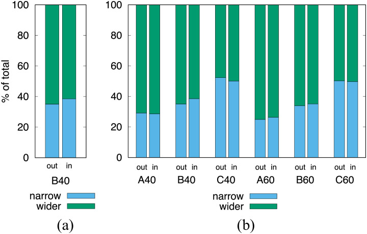
Fig 3. Percentage split of RBCs as they enter the daughter branches.
(a) The out column shows the situation when the rare cell is in the parent channel and the in column when the rare cell is in the daughter channel. (b) Comparison of geometries and hematocrits. In the C cases, where both daughter branches have equal width, the one reported as wider is the one the rare cell entered.

