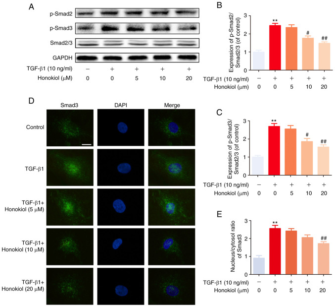
Figure 4.
Effect of honokiol on TGF-β1-induced Smad2/3 pathway activation. (A) Fibroblasts were pre-treated with honokiol for 2 h, and TGF-β1 was added for an additional 1 h. The cell lysates were then examined for the expression of p-Smad2, p-Smad3, and Smad2/3 by western blot analysis. (B and C) The ratios of the intensity of p-Smad2 and p-Smad3 relative to Smad2/3. (D) Fibroblasts were treated with TGF-β1 and/or various concentrations (5, 10 or 20 µM) of honokiol for 2 h prior to confocal microscopy (scale bar, 10 µm). (E) The nuclear/cytosolic ratio of Smad 3 staining was measured. Data are presented as the means ± SEM, **P<0.01, vs. the control group. #P<0.05, ##P<0.01 compared with the TGF-β1-stimulated group; n=3.

