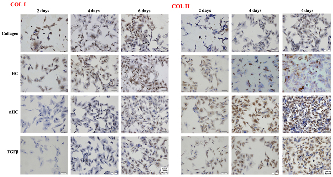
Figure 6.
Immunohistochemical staining. Stronger positive expression (brown-yellow color) of COL-II and weaker expression of COL-I was observed in the nHC group compared with that in the HC and pure collagen groups, indicating that the chondrcocyte phenotype and biological functions could be maintained effectively when cells were cultured on nHC film for a long time period. HC, hydroxyapatite/collagen; nHC, nano-HC; calcein-AM/PI, acetoxymethyl/propidium iodide; TGF-β, transforming growth factor-β; COL-I, collagen type I; COL-II, collagen type II.

