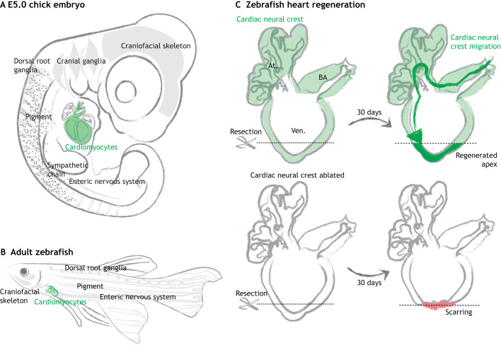
Fig. 2.
Neural crest-derived cardiomyocytes contribute to zebrafish heart regeneration. (A,B) Neural crest-derived cells persist as differentiated cells or stem cells residing in multiple organs, such as the craniofacial skeleton, peripheral nervous system, pigment of the skin (gray) and cardiovascular systems (green) in both amniotes (A) and zebrafish (B). (C) In the normal zebrafish heart, neural crest cells give rise to heart muscle (green) in the bulbus arteriosus (BA), atria (At.) and ventricles (Ven.). When the apex is surgically removed, these neural crest-derived cells participate in heart regeneration by migrating towards the injury site and redeploying genes that regulate neural crest development, such as Sox10. Genetic ablation of these cardiac neural crest cells disrupts heart regeneration, leading to scarring (red) (Tang et al., 2019b; Sande-Melón et al., 2019). Gray arrows represent regeneration period.

