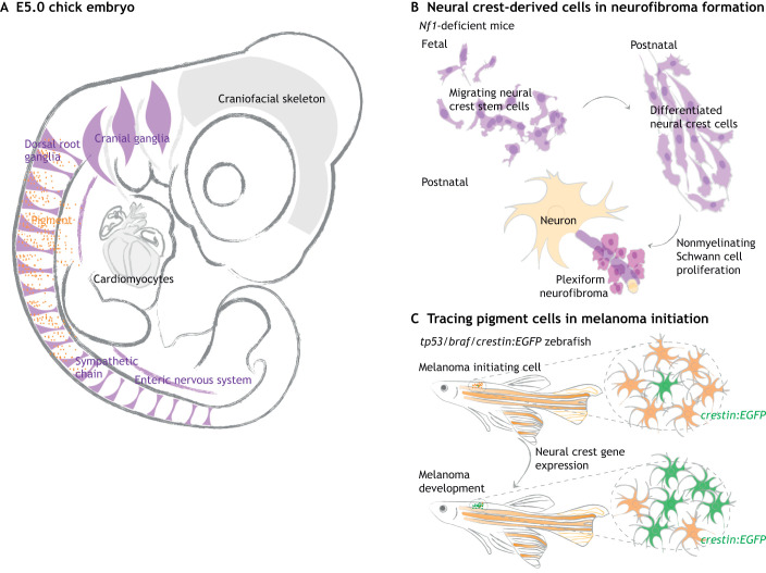
Fig. 3.
Tracing neural crest cells in tumor development. (A) Neural crest-derived cells contribute to the peripheral nervous system (PNS, purple) and pigment of the skin (orange), as illustrated in a chick embryo at E5. (B) Plexiform neurofibroma is a tumor of the PNS caused by mutations in the tumor suppressor gene Nf1. By tracing neural crest cells throughout the tumorigenesis process in Nf1 knockout mice, studies found that Nf1-deficient neural crest cells differentiate normally; tumors subsequently form by proliferation of differentiated Schwann cells. Thus, neural crest stem cells are not directly responsible for plexiform neurofibroma. (C) Neural crest lineage tracing can help identify early signs of melanoma. In a zebrafish model with oncogenic mutations in tp53 and braf, melanoma initiation is characterized by a normal pigment cell (orange) re-expressing neural crest-related genes, as visualized by crestin:EGFP (green). This particular cell transforms into a neural crest progenitor-like cell and later expands into a tumor. Gray arrows represent development.

