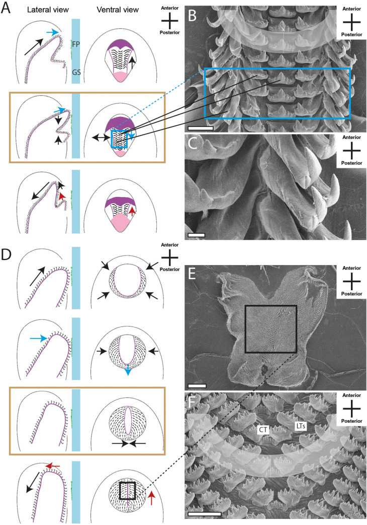
Fig. 6.
Schematic illustrations of radular motion patterns IV (A) and VI (D) in lateral and ventral view as seen through the glass surface. Blue arrow, motion in ventral direction; red arrow, motion in dorsal direction; black arrow, motion in horizontal/lateral direction; colored frame (blue, black) and black lines link these illustrations with SEM images of teeth (B,C, Marisa cornuarietis; E,F, Stenophysa marmorata). Brown frames emphasise the phase where two structures act as counter bearings. Scale bars: B,E, 200 μm; C, 40 μm; D, 20 μm.

