Fig. 5.
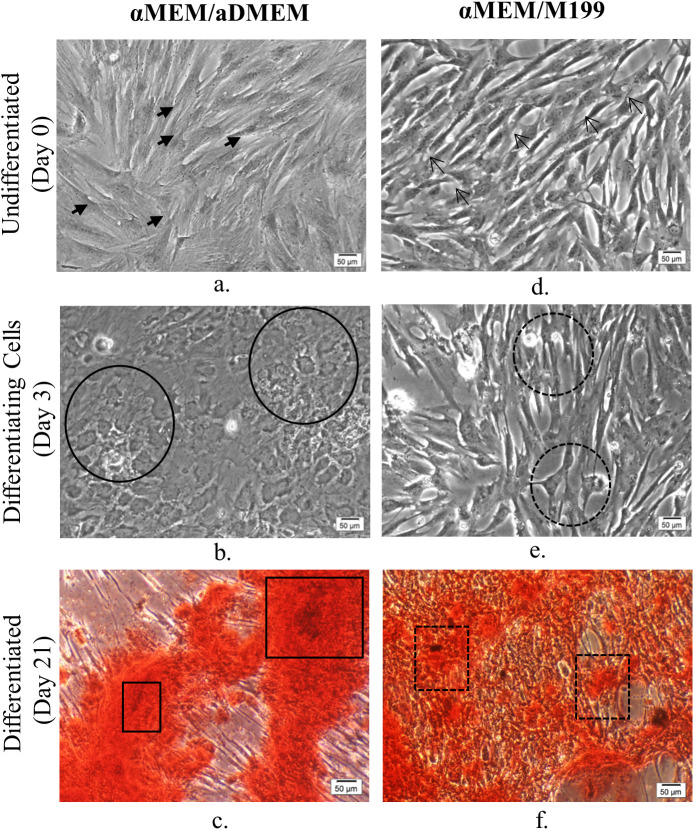
Phase-contrast micrographs of undifferentiated (day 0), osteogenically differentiating (day 3) and differentiated (day 21) passage 5 porcine bone marrow-derived MSC. αMEM/aDMEM grown undifferentiated cells (a) showing larger cell size (closed arrows) compared to spindle-shaped cells in αMEM/M199 (c) a typical of MSC morphology (opened arrows showing representative cells). The αMEM/aDMEM grown cells responded to osteogenic differentiation induction by changed shape to cuboidal morphology (line circles) as early as by day 3 (b) of differentiation, whereas, the cells grown in αMEM/M199 (d) did not show appreciable change in morphology (dotted circles) on the same day. The calcification assessed by Alizarin Red S stain at 21 days of osteogenic differentiation displayed heavily stained Alizarin Red S positive areas in αMEM/aDMEM grown pMSC(e) (line rectangles) whereas the pMSC grown in αMEM/M199 (f) showed patches of undifferentiated fibroblast-like cells (dotted rectangles) with very few cells displaying mineral deposition. Magnifications of respective images are as denoted by the scale bar.

