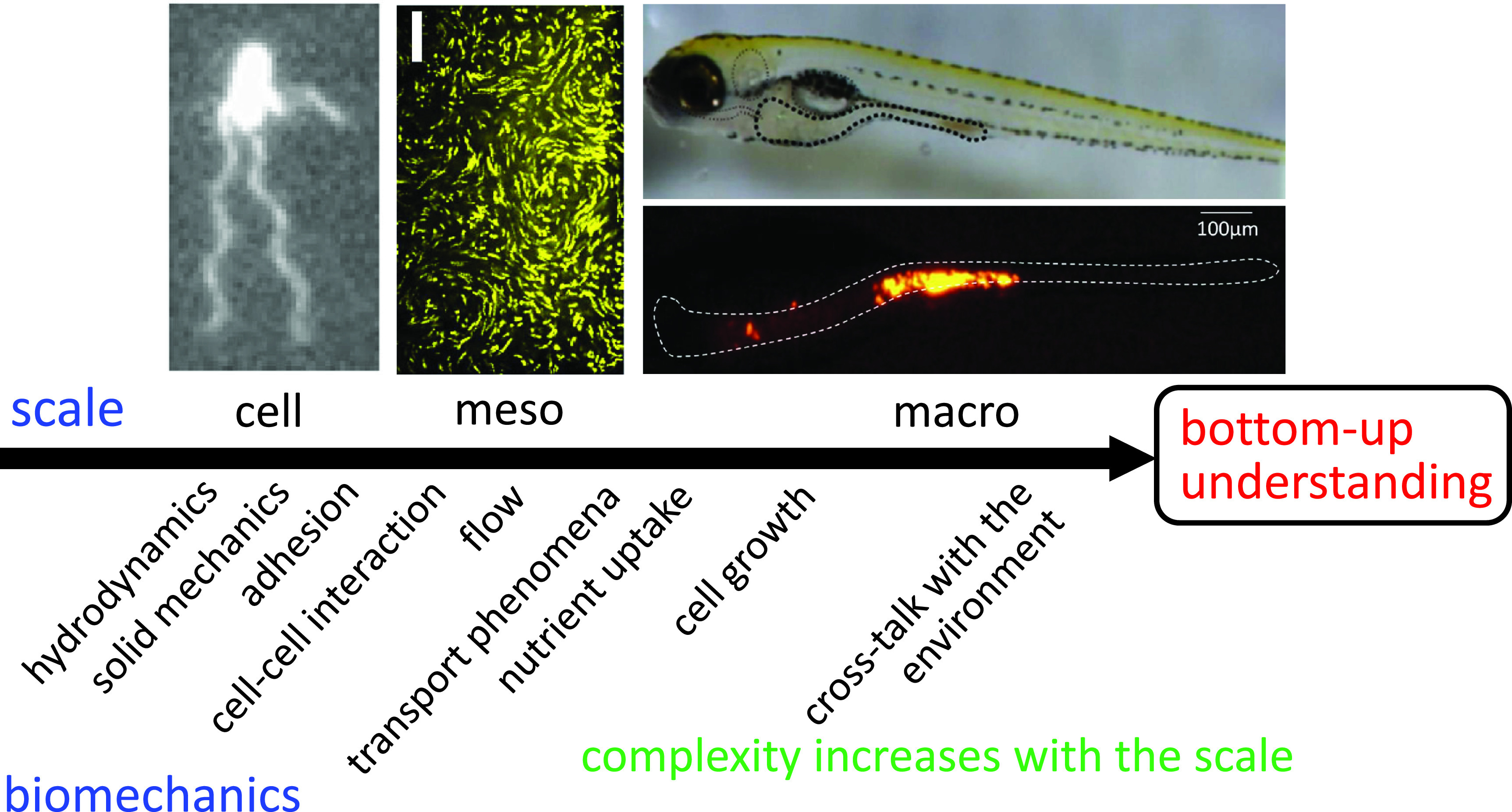
FIG. 1.

Schematic of a bottom-up understanding of bacterial biomechanics from the cellular to the macroscale. Top left, Escherichia coli cell; top middle, trajectories of tracers in a dense E. coli suspension; top right, zebrafish larva; and bottom right, fluorescent tracer particles in the intestine of zebrafish larva. The complexity of biomechanics increases with scale.
