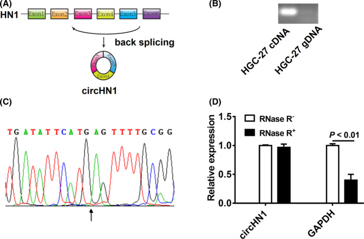
FIGURE 2.

Identification of circHN1. (A) Schematic drawing of the genomic location of circHN1. (B) PCR amplification of circHN1 with divergent primer by using cDNA and gDNA as the templates. (C) Back‐spliced junction sequence of circHN1 was validated by Sanger sequencing. (D) qRT‐PCR was performed to detect relative levels of circHN1 and GAPDH after treatment with (+) or without (−) RNase R
