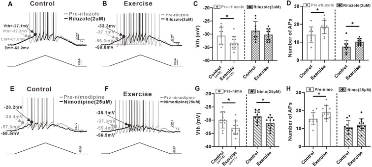
Figure 6.
Effects of PICs on neuronal excitability. (A) Example of riluzole (2 μM, black trace) reduced the firing frequency of 5-HT neuron compared to that of pre-riluzole (gray trace) and depolarized the Vth in control group. (B) Example of riluzole (2 μM, black trace) reduced the firing frequency of 5-HT neuron compared to that of pre-riluzole (gray trace) and depolarized the Vth in the exercise group. (C) Exercise training significantly depolarized the Vth (left panel, unpaired Student’s t-test, p = 0.044). After administration of riluzole, there was no significant difference in Vth between the control and exercise groups (right panel, unpaired Student’s t-test, p = 0.248). (D) Compared with the control group, exercise training increased the number of APs (left panel, unpaired Student’s t-test, p = 0.029). Even reduced by riluzole, the number of AP measured from the exercise group were still bigger than those from the control group (right panel, unpaired Student’s t-test, p = 0.034). (E) Example of nimodipine (25 μM) reduced the firing frequency of 5-HT neuron compared to that of pre-nimodipine (gray trace). (F) Example of nimodipine (25 μM) reduced the firing frequency of 5-HT neuron compared to that of pre-nimodipine (gray trace) in the exercise group. (G) Exercise training significantly hyperpolarized the Vth (left panel, unpaired Student’s t-test, p = 0.026). Significant difference was also observed in Vth between control and exercise groups with nimodipine (right panel, unpaired Student’s t-test, p = 0.013). (H) Compared with the control group, exercise training increased the number of APs (left panel, unpaired Student’s t-test, p = 0.025). After administration of nimodipine, there was no significant difference in the number of APs between control and exercise groups (right panel, unpaired Student’s t-test, p = 0.352). *p < 0.05.

