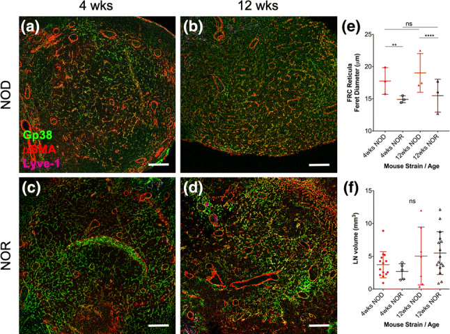
Figure 1.

FRC networks in NOD PLNs display larger/more relaxed reticular pores than NOR PLN reticula. (a–d|) PLNs were harvested from either female NOD (a, b) or female NOR (c, d) mice at 4 or 12 weeks of age and their volume was quantified; then, whole LNs were fixed in formalin, embedded in paraffin, sectioned at 5 µm, stained with antibodies against gp38 (identifies both FRCs and lymphatic endothelial cells), Lyve-1 (identifies only lymphatic endothelial cells), and αSMA (identifies myofibroblasts, including FRCs), and imaged using a confocal microscope. Shown are representative images of n = 3 mice. FRC reticula were identified as gp38+ (green), Lyve-1− (cyan) cells. Scale bars: 100 µm. (e, f) Quantification of reticular properties of FRC networks in PLNs as Feret’s diameter of gp38+ Lyve-1− FRC reticular pores (e) and overall PLN volume (f) of NOD PLNs compared to NOR PLNs from either 4-week old or 12-week old mice. Each data point represents the average of 30 pore quantifications per mice; n = 3 mice were analyzed. For quantification of LN volumes from LN photographs, biological replicates (mice) were n = 14 (4 weeks NOD), n = 5 (4 weeks NOR), n = 7 (12 weeks NOD), and n = 10 (12 weeks NOR). ns not significant, **p < 0.01, ****p < 0.0001.
