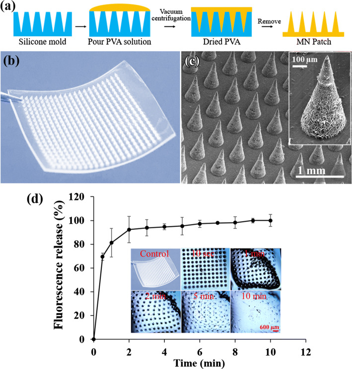
Figure 1.
(a) Fabrication of dissolvable MN patch using micromolding technique. (b) Photograph of dissolvable MN patch. (c) SEM images of the MN array (inset: single MN). The MN patch consist of a 21 × 21 needle array, and each MN has a 300 μm base width to prevent significant tissue damage and 600 μm length for penetration into the dermis. (d) Dissolution efficiency and RhB release profile from the MN patch in PBS (10 mL, pH 7.4) for up to 10 min at 37 °C. Each data point represents the mean RhB fluorescence intensity at 556 nm excitation and 580 nm emission spectrum peak wavelength. Inset of d shows the light microscopic images of MN patch captured at different time points.

