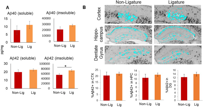
Figure 2.
Amyloid beta (Aβ) accumulation and topographical distribution in the cortex, hippocampal formation, and dentate gyrus. (A) Soluble and insoluble forms of Aβ in brain extracts from 5xFAD mice with (Lig) or without (Non-Lig) experimental PD. *p < 0.05 compared to non-PD animals. (B) Photomicrographs of Aβ42-immunostained sections containing cortical layers 4–5 (CTX), the hippocampus (HPC), and the dentate gyrus (DG) subregion of the hippocampus, were taken with a 4 × objective from Non-Lig and Lig 5xFAD mouse brain sections. A constant threshold was applied to each image. Analysis of all thresholded Aβ42 particles was performed to obtain the percent area of Aβ42 staining.

