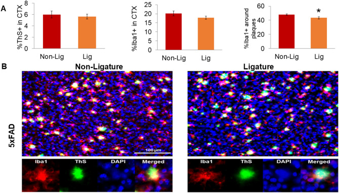
Figure 4.
Impact of ligature-induced PD on plaque-associated microglia (PAMs) in 5xFAD mice. Fluorescent photomicrographs containing Iba1+ microglia (red), thioflavin S (ThS) dense-core plaques (green), and DAPI cell nuclei (blue) were taken of cortical layers 4–5 (CTX) from Non-Lig and Lig 5xFAD mice (×20 objective, 2 images/section, 3 sections/animal). Each fluorescent channel was automatically thresholded; an ROI around each ThS+ plaque (including a 6.5 μm buffer-zone) was generated; and channels were analyzed within and without the ROI. (A) Dense-core plaque densitometry (%ThS+), microglia densitometry (%Iba1+), and the percentage of PAMs (%Iba1+ staining within the immediate proximity of ThS+ plaques), *p < 0.05. (B) Representative fluorescent photomicrographs of Non-Lig (left panels) and Lig (right panels) 5xFAD mouse cortex. Scale bar = 100 μm. Bottom row: Iba1, ThS, DAPI, and merged channels from a representative image of a PAM.

