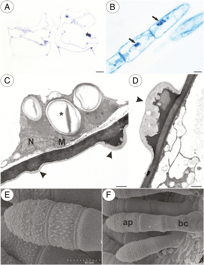
Fig. 2.
Structure of the non-glandular trichomes. (A) Section through the P. albida multicellular thick compact non-glandular trichomes that are located in the throat; note the numerous starch grains (asterisk); scale bar = 10 µm. (B) Naphthol blue black staining of a P. moctezumae multicellular non-glandular trichome showing the presence of a nucleus with a paracrystalline protein inclusion (arrow); note there are no protein bodies in the cytoplasm; scale bar = 10 µm. (C and D) Ultrastructure of a cell of a P. agnata non-glandular trichome; note the mitochondrion (M), nucleus (N) and prominent cuticular striations (arrowhead); scale bars = 0.7 µm and 0.5 µm, respectively. (E and F) Micromorphology of a P. agnata multicellular compact thick non-glandular trichome that is located in the front of the throat; note the cuticular striations on the surface of the apical cells (ap) and the smooth cuticle surface of the basal cell (bc); scale bars = 50 µm and 100 µm, respectively.

