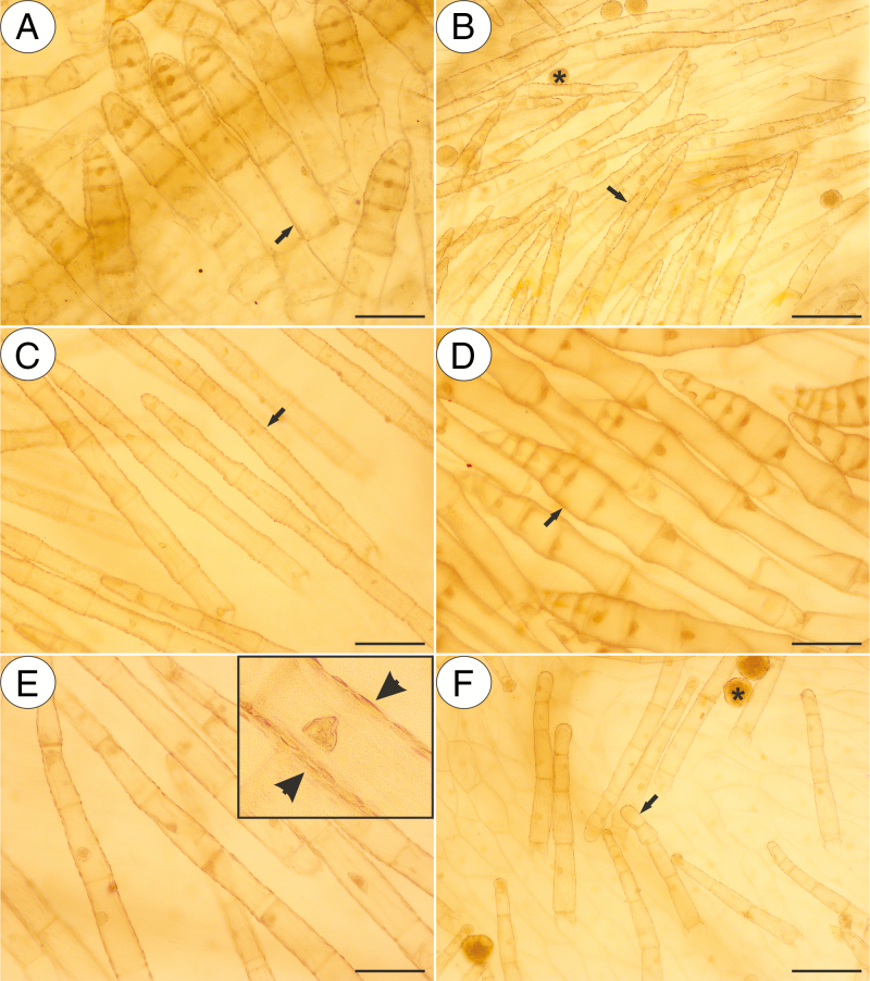
Fig. 9.
Sudan III staining of various non-glandular trichomes of the selected Pinguicula species that were examined; note the positive staining of the cuticular striations of the non-glandular trichomes cells (arrow, insert and arrowhead) and lipids inside the pollen grains (asterisk). (A) P. agnata; scale bar = 100 µm. (B) P. rectifolia; scale bar = 100 µm. (C) P. moranensis; scale bar = 100 µm. (D) P. esseriana; scale bar = 100 µm. (E) P. hemiepiphytica; scale bar = 100 µm. (F) P. mesophytica; scale bar = 100 µm.

