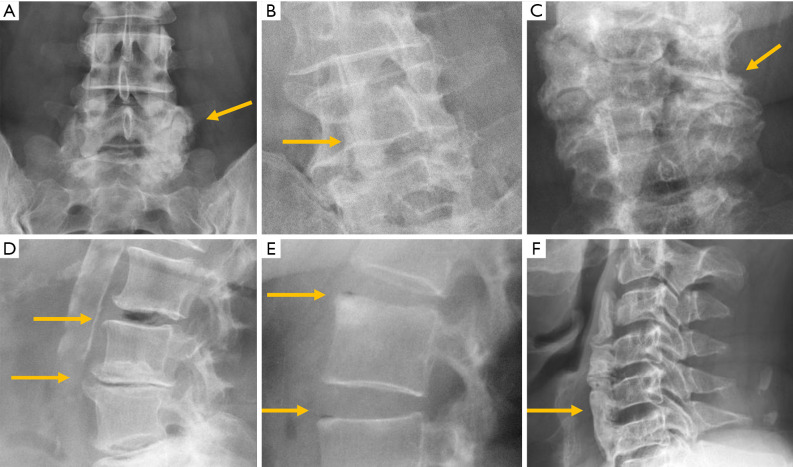
Figure 13.
Facet osteoarthritis. AP (A) and oblique (B) lumbar radiographs. (C) Facets osteoarthritis at the cervical spine; (D) vertebral osteochondrosis with marginal osteophytes and central vacuum phenomenon; (E) spondylosis deformans with marginal sclerosis and peripheral vacuum phenomenon; (F) diffuse idiopathic skeletal hyperostosis. Arrows point the main abnormality in each case.

