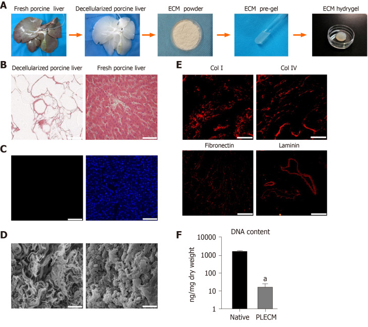
Figure 1.
Formation and characterization of porcine liver extracellular matrix gel. A: Procedure for the formation of porcine liver extracellular matrix (PLECM) gels; B: Hematoxylin and eosin (HE) staining of PLECM (left) and porcine liver (right). Scale bars = 100 μm; C: 4,6-diamidino-2-phenylindole staining of PLECM (left) and porcine liver (right). Scale bars = 100 μm; D: Scanning electron microscopy of PLECM (left) and porcine liver (right). Scale bars = 20 μm; E: Immunohistochemistry (red) for liver extracellular matrix proteins (collagen type I, collagen type IV, fibronectin, and laminin) in PLECM. Scale bars = 200 μm; F: Relative DNA content. aP < 0.05 vs PLECM. ECM: Extracellular matrix; Col I: Collagen type I; Col IV: Collagen type IV; PLECM: Porcine liver extracellular matrix.

