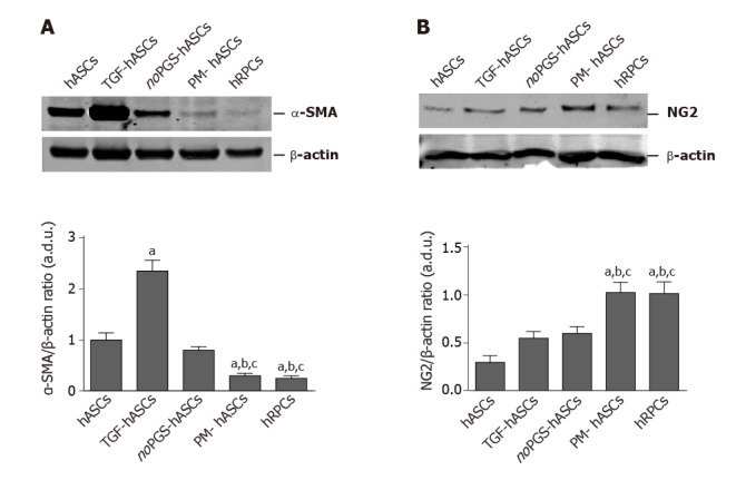
Figure 3.

Western blot analysis of alpha smooth muscle actin and neural/glial antigen 2 expression in different groups of human adipose-derived mesenchymal stem cells and human retinal pericyte cells at 3 d of culture. A: Western blot analysis of alpha smooth muscle actin expression; B: Western blot analysis of neural/glial antigen 2 expression. Histograms in A show that alpha smooth muscle actin levels measured in control human adipose-derived mesenchymal stem cells pre-cultured in basal medium (hASCs) are similar to hASCs pre-cultured in pericyte medium lacking pericyte growth supplement (noPGS-hASCs); much higher levels were found in hASCs pre-stimulated with transforming growth factor (TGF-hASCs); the lowest levels were observed in hASCs pre-cultured in complete pericyte medium (PM-hASCs), close to those for human retinal pericyte cells (hRPCs). Histograms in B show that neural/glial antigen 2 levels measured in control hASCs are slightly higher in TGF-hASCs and noPGS-hASCs; significant increases were observed in PM-hASCs, similar to those for hRPCs. All data represent mean ± SEM obtained from at least three independent experiments. Comparison between groups was evaluated by one-way ANOVA, followed by Tukey’s test. aIndicates significant difference (P < 0.05) vs hASCs; bIndicates significant difference (P < 0.05) vs TGF-hASCs; cIndicates significant difference (P < 0.05) vs noPGS-hASCs. α-SMA: Alpha smooth muscle actin; hASCs: Human adipose-derived mesenchymal stem cells pre-cultured in basal medium; hRPCs: Human retinal pericyte cells; NG2: Neural/glial antigen 2; noPGS-hASCs: hASCs pre-cultured in pericyte medium lacking pericyte growth supplement; PM-hASCs: hASCs pre-cultured in complete pericyte medium; TGF-hASCs: hASCs pre-stimulated with transforming growth factor.
