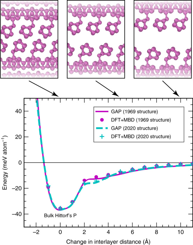
Fig. 4. Exfoliation of Hittorf’s phosphorus.

Exfoliation into monolayer “hittorfene”73, similar to Fig. 2d, but now for a more complex structure where training data are only available around the minimum. Two different experimental structural models are used as a starting point: the initial P2/c structure (1969, ref. 9, magenta), and a very recently proposed P2/n structure (2020, ref. 11, cyan). The results of our GAP + R6 model are given by solid and dashed lines, respectively, and reference DFT + MBD computations are indicated by circles and crosses.
