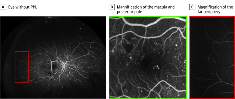
Figure 1. Distribution of Diabetic Retinopathy Lesions in an Example Eye Without Predominantly Peripheral Lesions (PPL).
A, Eye without PPL. B, Magnification of the macula and posterior pole. C, Magnification of the far periphery. Most of the lesions are localized to the macula and posterior pole (green) with minimal microaneurysms present in the far periphery (red).

