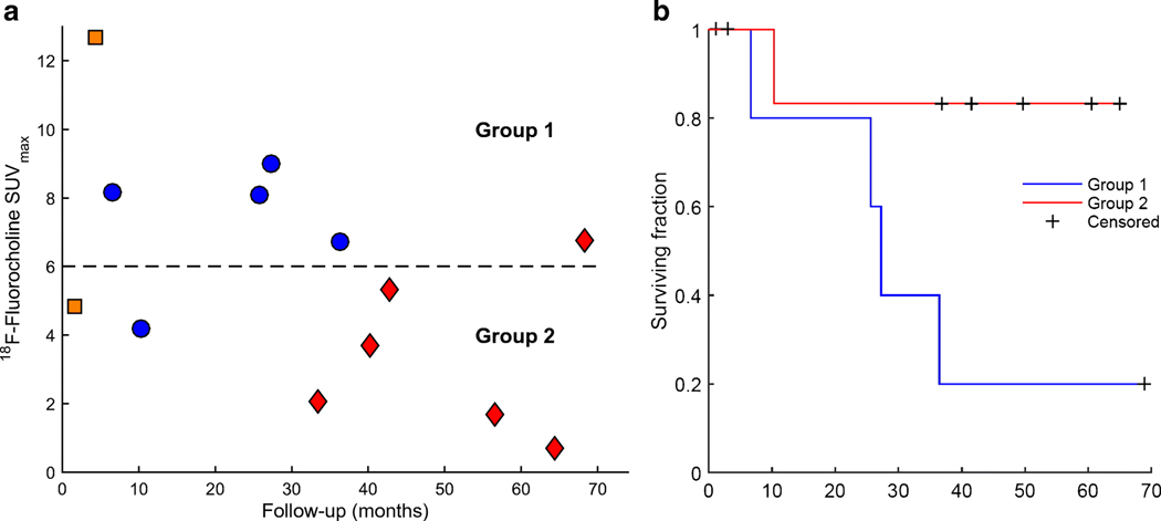
Figure 3.
(A) 18F-Fluorocholine PET SUVmax as a function of follow-up time for n=13 pts (follow-up was not established for one international patient). Blue circles - deceased from previously treated progressing brain metastases (n=5). Orange square - deceased from progressing metastases in the liver and pancreas (n=1) and previously untreated brain metastases (n=1). Red diamonds - alive (n=6). Dashed line represents 18F-Fluorocholine PET SUVmax = 6 (median value), splitting the data into two groups. (B) Kaplan-Meier estimator for groups 1 and 2. Observations were censored if the patient was alive at the time of last follow-up or deceased from cause other than progression in the treated brain metastases. Difference in survival distributions did not reach significance according to the log-rank test (p=0.068).

