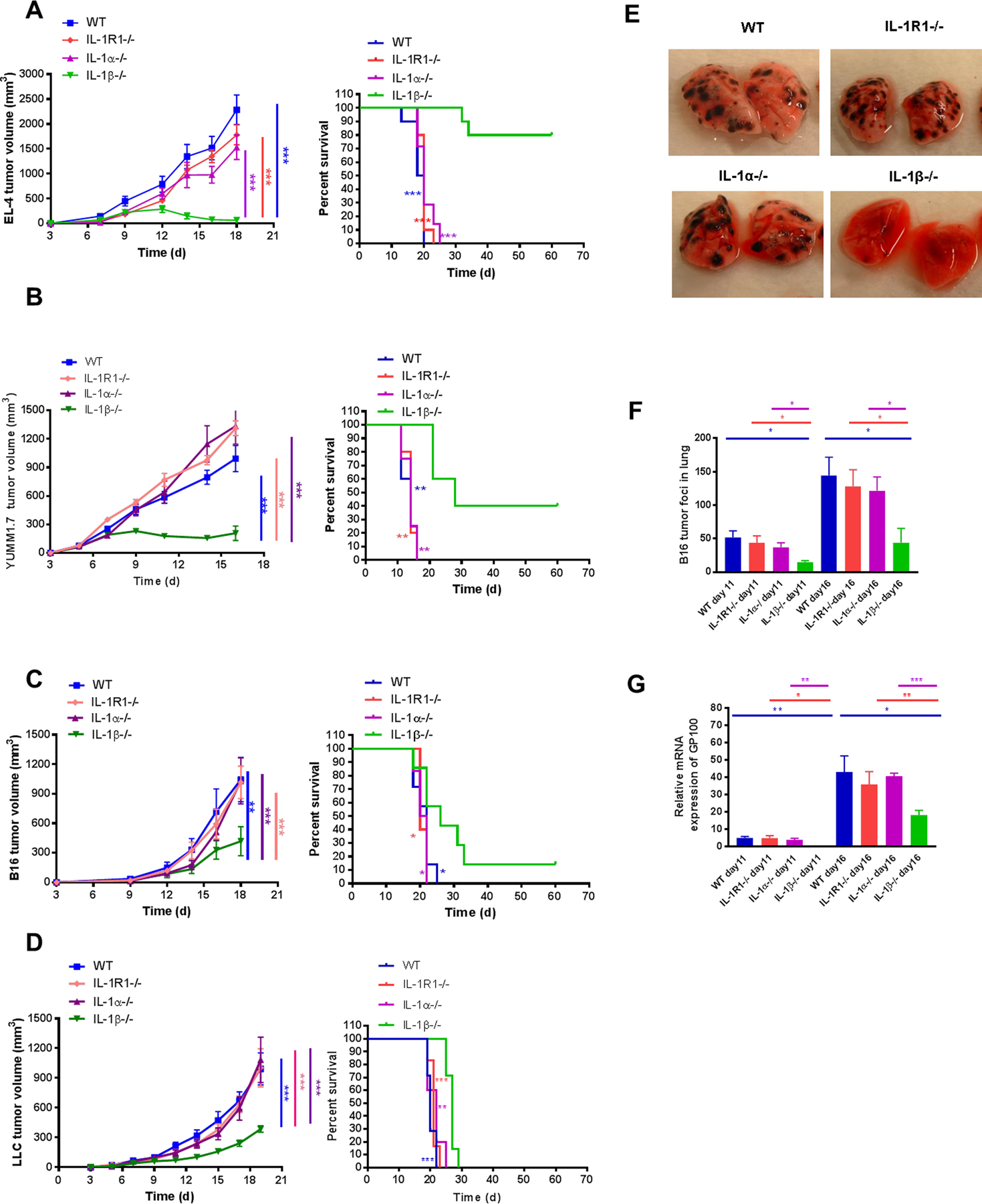
Figure 1. IL1β−/− mice, but not IL1α−/− or IL1R1−/− mice demonstrated reduced tumor growth in skin and tumor metastasis in the lung.

(A) EL4 lymphoma (2×10⁵), (B) YUMM1.7 melanoma (3×10⁶), (C) B16-F10 melanoma (3×10⁵), and (D) LLC (2×10⁵) cells were injected i.d. into WT and IL1-deficient mice. Tumor growth (left) and survival (right) were recorded every 2–3 days and statistics generated as compared to IL1β−/− mice (n=5–10 mice per group). B16F10 melanoma cells (5×10⁵) were injected i.v. into WT and IL1-deficient mice. Tumor foci in the lungs were (E) photographed at day 16 and (F) counted at day 11 and 16 (n=4–5 mice per group). (G) Expression of GP100 in lungs of WT and IL1-deficient mice was assessed using qPCR. GP100 was quantified relative to the housekeeping gene GAPDH. Relative mRNA expression of GP100 was normalized to control WT mice set at 1. Graphs show mean ± SEM. *P < 0.05, **P < 0.01, ***P < 0.001. NS=not significant.
