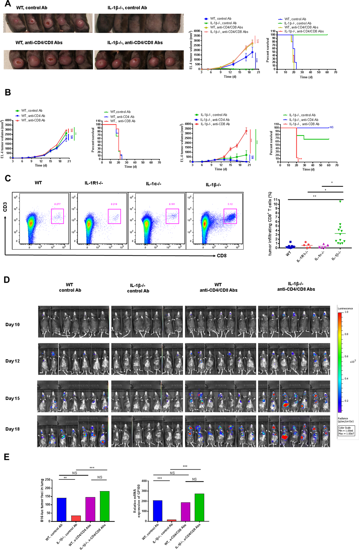
Figure 3. EL-4 tumor growth and B16 melanoma metastasis were restored in IL1β−/− mice after T-cell depletion.

(A) 2×10⁵ EL-4 cells were injected i.d. into WT and IL1β−/− mice treated with anti-CD4/CD8 or control Abs 2 days before and then every 2–3 days after tumor cell injection. Tumors were photographed at day 17 after EL-4 injection (left) and tumor volumes (middle) were plotted. Survival was also recorded (right) (n=5 mice per group). (B) 2×10⁵ EL-4 cells were injected i.d. into IL1β−/− and WT mice treated with either anti-CD4, anti-CD8 or control Ab. Tumor growth and survival was recorded every 2–3 days and plotted. (n=8–9 mice per group). (C) 2×10⁵ EL-4 cells were injected i.d. into WT and IL1 deficient mice. Representative dot plots were shown and percentage of tumor infiltrating CD8+CD3+ T cells were measured at day 14 after EL-4 cell injection (n=5–12 mice per group). (D) 5×10⁵ B16-F10 cells expressing luciferase (B16-Luc) were injected i.v. into WT and IL1β−/− mice treated with either control Ab or antibodies to both CD4 and CD8 2 days before and then every 2–3 days after tumor cell injection. Bioluminescence imaging was performed at different time points (n=7 mice per group). (E) Tumor foci in the lungs were numerated and the expression of GP100 was measured at day 19 after B16-Luc i.v. injection (n=7 mice per group).
