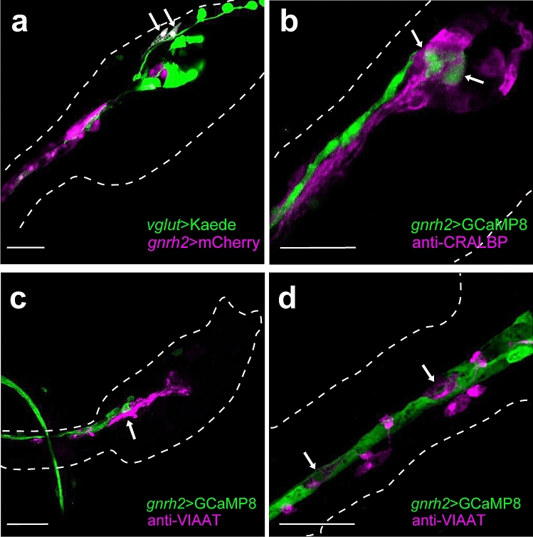
Figure 2.
Immunohistochemical identification of types of cells expressing gnrh2 in the Ciona larva at 21 hpf. (a) Glutamatergic neurons and gnrh2-expressing cells were labeled with Kaede (green) and mCherry (magenta), respectively. The proto-placode-derived sensory neurons (aATENs; arrows) were shown to express gnrh2 (6 of 6 larvae showed the expression; four additional examples are shown in Supplementary Figure S2). (b) CRALBP-positive cells (magenta) were not overlapped with gnrh2-expressing cells (green) (none of 16 larvae showed overlapped expression; four additional examples are shown in Supplementary Figure S3). Arrows indicate gnrh2-expressing cells in the brain vesicle. (c, d) GABAergic/glycinergic neurons were visualized by immunostaining with anti-VIAAT antibody (magenta). VIAAT-positive cells (magenta) were not overlapped with gnrh2-expressing cells (green) (none of 17 larvae showed overlapped expression; four additional examples are shown in Supplementary Figure S3). Arrows in (c) indicate GABAergic/glycinergic neurons in the motor ganglion. Arrows in (d) indicate VIAAT-positive ACINs. (a) Projection of 7 serial optical sections taken at 0.60 µm intervals. (b) Projection of 8 serial optical sections taken at 0.60 µm intervals. (c) Projection of 7 serial optical sections taken at 0.60 µm intervals. (d) Projection of 3 serial optical sections taken at 0.60 µm intervals. Scale bars, 30 µm.

