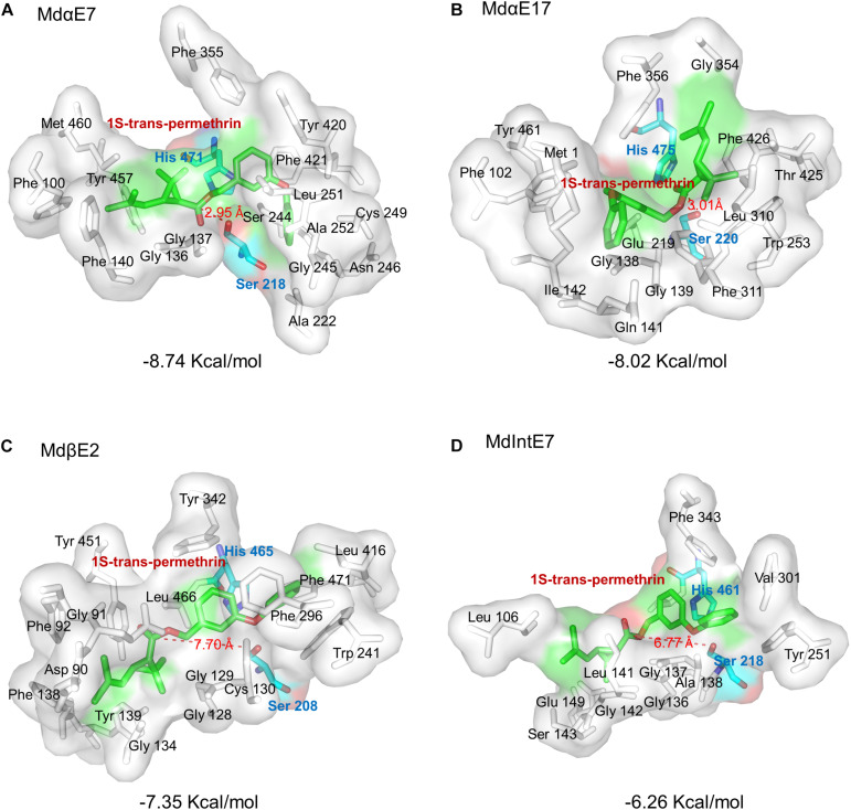
FIGURE 4.
Stereo view of permethrin bound within the active site cavity. Permethrin bound within the active site cavity of: (A) MdαE7; (B) MdαE17; (C) MdβE2; and (D) MdIntE7. The Serine (Ser) and Histidine (His) residues are labeled as green sticks and the 1S-trans-permethrin isomer as cyan sticks. The distance between permethrin and the Ser residue of the carboxylesterase is indicated by a red dashed line. The binding energy is also shown. All amino acid active site cavities are labeled in each carboxylesterase structure.

