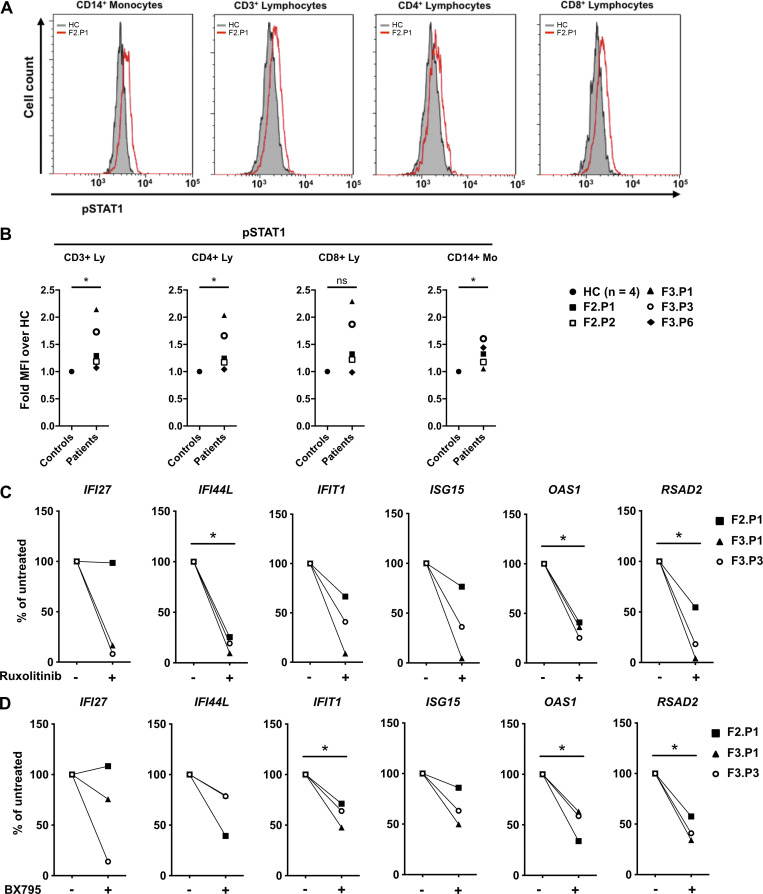
Figure 2.
Type I IFN is responsible for the constitutive induction of ISGs in COPA syndrome. (A) Flow cytometry analysis of phosphorylated STAT1 (pSTAT1) in monocytes, CD3+, CD4+, and CD8+ lymphocytes from F2.P1 compared with a HC. (B) pSTAT1 measured as in A in CD3+, CD4+, and CD8+ lymphocytes (Ly) and CD14+ monocytes (Mo) from COPA patients compared with a HC sampled the same day, and expressed as fold mean fluorescence intensity (MFI) of over that of the HC. Blood from F3.P1 and F3.P3 was processed during the same experiment and compared with one HC. Data were statistically analyzed using the Mann–Whitney test (*, P < 0.05; ns, not significant). (C and D) RT-qPCR gene expression analysis in PBMCs from three patients (F2.P1, F3.P1, and F3.P3), after overnight culture in the absence of treatment or presence of ruxolitinib (C) or BX795 (D). Results are expressed as percentage of the baseline elevated levels and show that IFIT1, IFI27, IFI44L, ISG15, OAS1, and RSAD2 levels decreased after in vitro treatment (except IFI27 in F2.P1) for both drugs. Data were statistically analyzed using a one-sample t test (*, P < 0.05).

