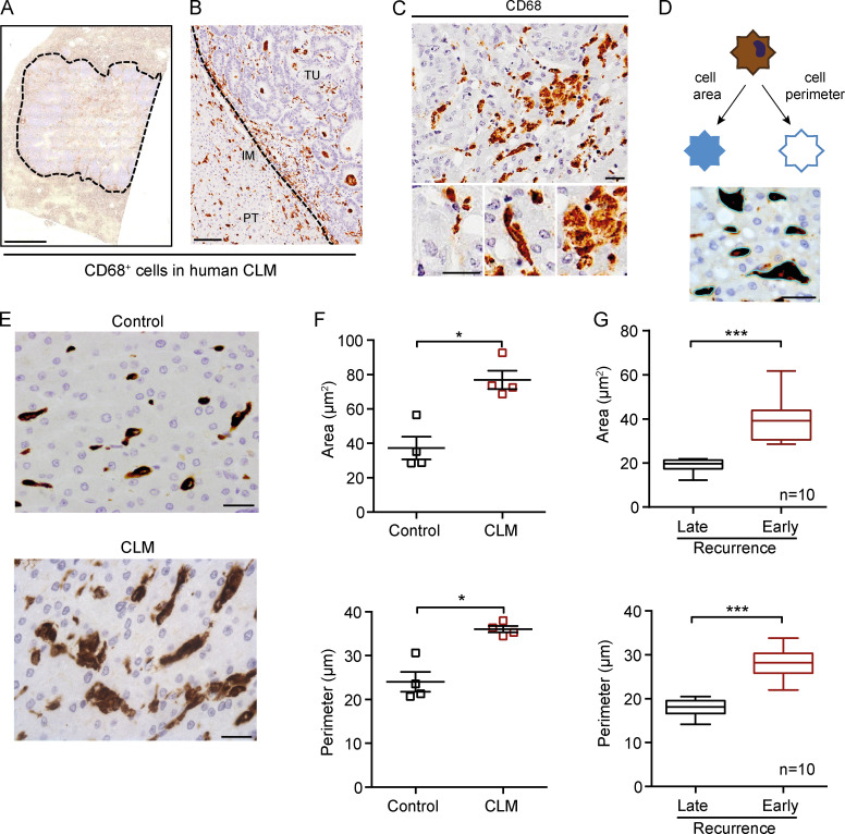
Figure 1.
Morphological assessment of macrophages in human CLMs. (A) Representative whole slide immunohistochemistry of CD68+ cells in a CLM specimen. Dotted line indicates the tumor lesion. (B) CD68+ macrophages infiltrate both the PT area, IM, and tumor (TU) of CLM tissues. (C) Morphological features of macrophages in CLM. Three exemplificative types of macrophages in the same region are shown: small and round shaped, spiky and elongated, and big pancake-like. (D) Representative scheme of morphological features of macrophages analyzed (area and perimeter). Picture depicts the image analysis procedure. (E) Representative pictures of CD68+ macrophages in a control liver (symptomatic giant liver hemangioma, top) and a CLM specimen (bottom). (F) Both macrophage area and perimeter are significantly higher in CLM specimens compared with controls (i.e., patients who had undergone surgery for symptomatic giant liver hemangiomas). Represented are mean ± SEM of three pictures from each specimen (n = 4 specimens each group; *, P = 0.029 [area] and P = 0.028 [perimeter] by Mann-Whitney test). (G) Quantitation of macrophage area and perimeter in CLM specimens from patients with late recurrence (DFS >24 mo) or early recurrence (DFS <24 mo). Box plots give median, lower, and upper quartile values by the box and minimum and maximum values by the whiskers from three pictures for each specimen (n = 10 specimens each group; ***, P < 0.001 by unpaired t test). Scale bars: 2 mm (A), 100 µm (B), and 50 µm (C–E).

