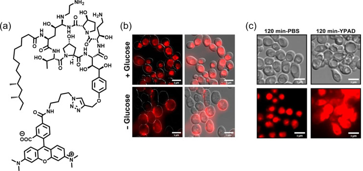
Figure 1.
(a) TMR-labeled caspofungin probe 1. (b) Microscopy images of C. albicans cells treated with probe 1 (red) in the presence/absence of glucose, indicative of endocytosis. (c) DIC and fluorescent images of C. albicans remaining viable in PBS (left) but displaying fungal cell death characteristics when treated with probe 1 in YPAD media (right) after 120 min. Reproduced with permission from ref (2). Copyright 2020 American Chemical Society.

