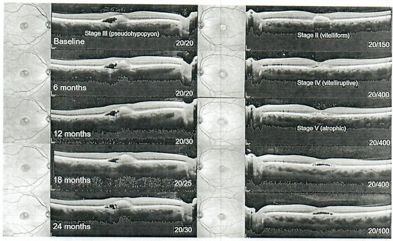
Fig. 6.

Serial spectral-domain optical coherence tomography imaging from baseline (pre-sildenafil) to 24 months in 6-month intervals in Best disease (patient 5). The right eye exhibited a pseudohypopyon (stage III) vilelliform lesion at presentation which remained stable over time, while the left eye initially presented at stage II and progressed to the atrophic stage after 1 year with an accompanying decline in visual acuity from 20/150 to 20/400. Visual acuity subsequently recovered to 20/100 by the second year of follow-up.
