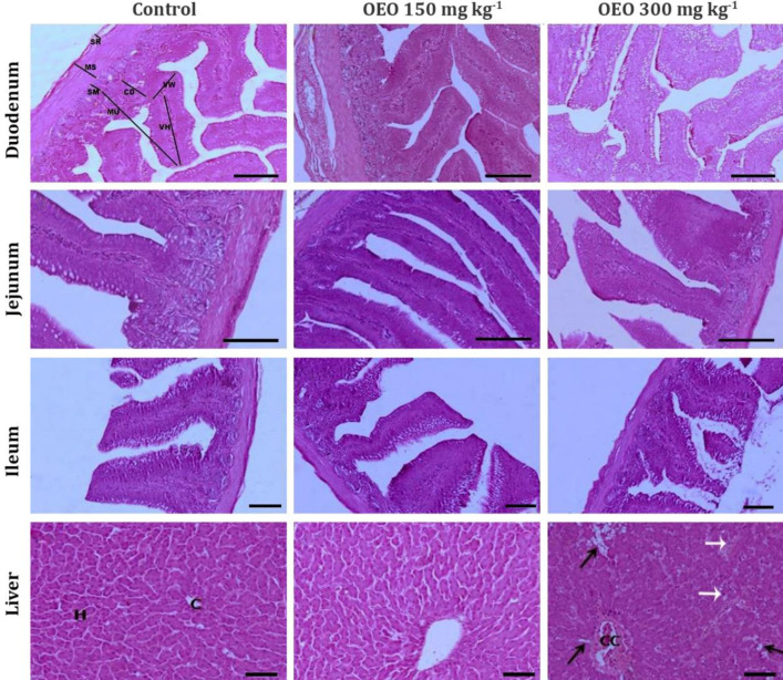
Fig. 1.
Light microscopic photomicrographs showed morphological characteristics of the parameters evaluated in particular regions of the quail alimentary tract. OEO: Oregano essential oil, VH: Villus height, VW: Villus width was near the crypt, CD: Crypt depth, MU: Thickness of intestinal mucosa, SM: Submucosa, MS: Tunica musculosa, and SR: Tunica serosa. H: Hepatocytes, C: Central vein, CC: Congested, and dilated central vein. Black arrows indicate fatty degeneration of hepatocytes, and white arrows show Kupffer cell hypertrophy, (H & E, Scale bar = 50 μm).

