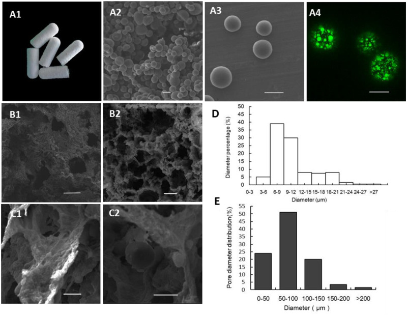
Figure 2.
Morphology of AMP@PLGA microspheres and AMP@PLGA/MC scaffolds. (A1) AMP@PLGA/MC scaffold. (A2, A3) Morphology of AMP@PLGA microspheres. (A4) The distribution of KSL-W (FITC) in PLGA microspheres. (B1, B2) The porous structure of MC scaffold. (C1, C2) MC and AMP@PLGA microspheres on scaffolds. (D) The diameter of AMP@PLGA microspheres. (E) The pore size of AMP@PLGA/MC scaffolds (A1–A3, C1 and C2 and B4 on a scale of 10 µm, B1 and B2 on a scale of 100 µm).

