Figure 6.
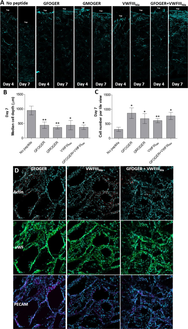
HUVEC Distribution in collagen scaffolds. HUVECs were seeded onto the top surface of EDC/NHS cross-linked scaffolds without peptides or with GFOGER, GMOGER, VWFIIINle or GFOGER + VWFIIINle, cultured for 4 or 7 days, fixed and stained with Hoechst 33342 (blue). (A) Representative tile views of scaffold cross-sections after 4 or 7 days of culture. Without peptides, HUVECs migrated into the scaffolds away from the seeding site. With THPs, HUVECs densely populated the seeding site at the top of scaffolds, with little migration and instead proliferated into adjacent pores. (B) Median cell depth after 7 days. Significance for each condition compared with cross-linked scaffolds without peptide is shown. Cell distribution was homogeneous on scaffolds without THPs, whereas most cells were located within the top half of the THP-functionalized scaffolds. (C) Cell number in cross-sections. Significance for each condition compared with cross-linked scaffolds without peptide is shown. Cell number was maintained over 7 days in THP-functionalized scaffolds and was significantly higher than scaffolds without peptides. (D) PECAM-1 (purple) and VWF (green) staining in HUVEC cultures on EDC/NHS cross-linked scaffolds functionalized with GFOGER, VWFIIINle or GFOGER + VWFIIINle. Representative Z-stacks from plan views of the top 30 µm of scaffolds are shown. PECAM-1 and VWF were detected in all cells, indicating that HUVECs cultured for 7 days on cross-linked scaffolds maintained an endothelial phenotype.

