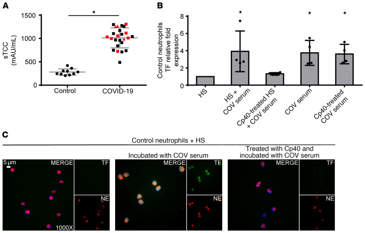
Figure 3. C3 inhibition disrupts neutrophil-driven thromboinflammation in COVID-19.
(A) Soluble terminal complement complex (sTCC) levels in plasma from controls (n = 10) and patients with COVID-19 (n = 25). Red squares: severe patients; red triangles: critical patients. (B) Relative fold expression of TF mRNA in control neutrophils stimulated with serum from healthy individuals (HS), HS incubated with COVID-19 serum (COV serum), or HS treated with compstatin analog Cp40 and then COV serum, COV serum alone, or COV serum treated with Cp40. Data are from 4 independent experiments (mean ± SD). (C) Fluorescence microscopy showing TF/NE staining in control neutrophils stimulated with HS, HS incubated with COV serum, or HS treated with Cp40 and then COV serum. A representative example of 4 independent experiments is shown. Original magnification: ×1000; scale bar: 5 μm. Blue: DAPI, green: TF, red: NE. All conditions were compared with HS alone (control). *Statistical significance at P < 0.05. A: Student’s t test, B: Friedman’s test. These in vitro experiments are also listed in Supplemental Table 2. Conditions of real-time RT-PCR are described in Supplemental Table 3.

