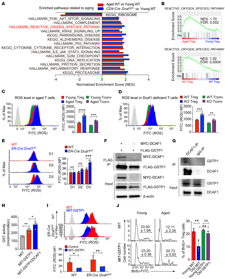
Figure 7. DCAF1 is required to suppress ROS in Tregs.
(A) Pathways commonly enriched in aged and Dcaf1-deficient (CD4-Cre Dcaf1fl/fl) Tregs based on GSEA of RNA-Seq data sets (FDR < 0.05). (B) Enrichment of ROS pathway in aged versus young WT Tregs (top) and Dcaf1-deficient (KO) versus WT Tregs (bottom) by GSEA of RNA-Seq data sets. (C–E) Flow cytometry of ROS level in indicated T cell populations from young WT and aged WT mice (C), young Dcaf1-deficient mice (D) and activated WT and ER-Cre Dcaf1fl/fl CD4+ T cells treated with 4-hydroxy-tamoxifen for indicated days (E), analyzed by DCFDA (gray area, no DCFDA; n = 3 mice of 3 experiments; representative results are shown; means ± SD, **P < 0.01, ***P < 0.001, ****P < 0.0001, by 2-way ANOVA followed by Holm-Šidák multiple-comparisons test). (F and G) Interaction of GSTP1 and DCAF1 by coimmunoprecipitation in 293T cells (F) and by endogenous immunoprecipitation using anti-DCAF1 antibody in mouse T cells (G). The results are representative of 3 independent experiments. (H) GST activity in 293T cells after overexpression of GSTP1 and DCAF1 for 4 days; n = 5; means ± SD, *P < 0.05, **P < 0.01, by Mann-Whitney U test. (I) Flow cytometry of ROS level in activated WT and ER-Cre Dcaf1fl/fl CD4+ T cells transduced with MIT (MSCV-IRES-Thy1.1) or MIT-GSTP1 virus in the presence of 4-hydroxy-tamoxifen, analyzed by DCFDA (gray area, no DCFDA; n = 3–4 experiments; means ± SD, *P < 0.01, **P < 0.05, by 2-way ANOVA followed by Holm-Šidák multiple-comparisons test). (J) Proliferation assayed by BrdU incorporation in young and aged Tregs transduced with MIT or MIT-Gstp1 virus (n = 3 experiments; means ± SD, **P < 0.01, by 1-way ANOVA followed by Tukey’s multiple-comparisons test).

