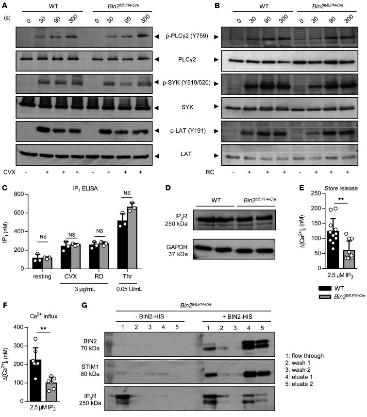
Figure 3. Defective IP3R function in Bin2fl/fl,Pf4Cre mice and BIN2 interaction with STIM1 and IP3R.
(A and B) Determination of whole-cell tyrosine phosphorylation pattern in WT and Bin2fl/fl,Pf4Cre platelets upon activation with convulxin (CVX) (A) or rhodocytin (RC) (B). The samples were taken at the indicated time points, lysed, and Western blot analysis was performed with the indicated phospho-specific and pan-antibodies. This blot is representative of 3 independent experiments. (C) Quantification of inositol monophosphate (IP1), a specific metabolite of inositol-1,4,5-trisphosphate (IP3), produced upon activation with the indicated agonist (n = 3). Representative results of 3 independent experiments. (D) Western blot analysis of IP3R expression in WT and Bin2fl/fl,Pf4Cre platelet lysates; GAPDH was used as loading control. (E and F) Ca2+ concentrations in the cytoplasm of WT and Bin2fl/fl,Pf4Cre platelets upon treatment with UV light–inducible IP3 in the (E) absence or (F) presence of extracellular Ca2+ (n = 8). (G) Bin2fl/fl,Pf4Cre platelet lysates were incubated with recombinant BIN2-HIS protein, followed by a purification step with NI-NTA beads. The different fractions were eluted and analyzed by Western blotting using BIN2-, STIM1-, and IP3R-specific antibodies. Representative result of 3 independent experiments. Values are depicted as mean ± SD, and P values were calculated using the Mann-Whitney U test. **P < 0.01. See complete unedited blots in the supplemental material.

