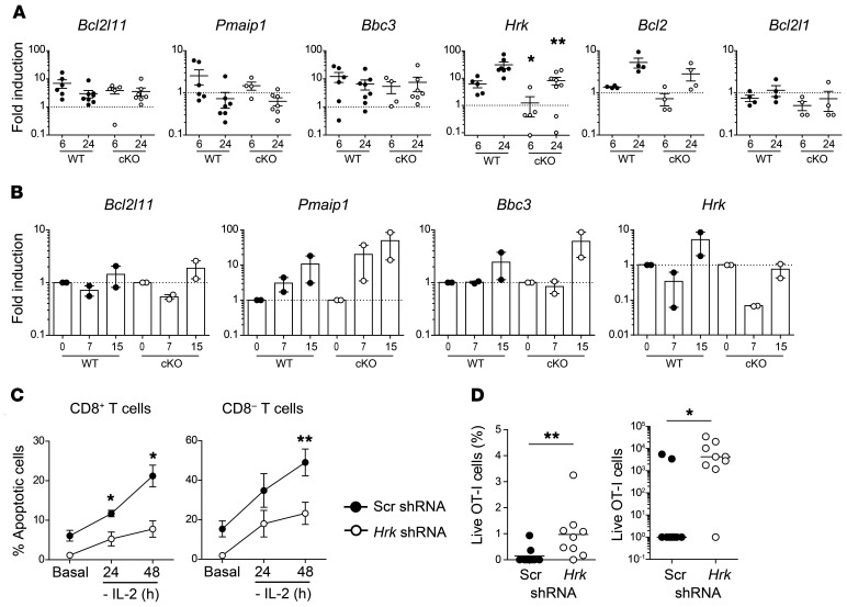
Figure 6. B55β-induced Hrk is essential for CWID.
(A) T cells from Ppp2r2b+/+Cd4.Cre+ (WT) or Ppp2r2bfl/flCd4.Cre+ (cKO) mice were activated and expanded in vitro. Expression of the indicated genes was assessed by qPCR before (basal) and after 6 and 24 hours of cytokine withdrawal. Cumulative data from 4 experiments (n = 3–4 mice/group/experiment) are shown. Mean and SEM are indicated. *P ≤ 0.05, **P ≤ 0.01 (unpaired 2-tailed Mann-Whitney test). (B) WT or cKO OT-I cells were adoptively transferred into CD45.1+ mice. Next day, LM-OVA was injected into recipient mice. RNA was extracted from sorted cells (CD45.2+ CD8+ Vα2+ Vβ5+) at days 7 and 15 and qPCR was performed. Results are presented as mean and SEM. Cumulative data from 2 independent experiments (n = 3–5 mice/group/experiment) are shown. In A and B, the dotted line indicates the expression level of the basal sample. (C) T cells from WT mice were activated and infected with control (Scr) or Hrk-shRNA encoding lentiviruses. Cells were expanded and apoptosis was induced by cytokine withdrawal. GFP+ (effectively infected) apoptotic cells were quantified. Cumulative data from 2 experiments (n = 4 mice/group/experiment). *P ≤ 0.05, **P ≤ 0.01 (unpaired 2-tailed t test). (D) CTLs from OT-I WT or cKO mice were infected with a control (Scr-Cerulean) or an Hrk-specific shRNA encoding lentivirus (GFP). Six days later, 1.6 × 10⁴ Cerulean+ and GFP+ OT-I T cells were injected at a 1:1 ratio into RIP-mOVA mice. Fourteen days later, relative and absolute numbers of transferred cells (Cerulean or GFP+) were quantified (n = 9). *P ≤ 0.05, **P ≤ 0.01 (unpaired 2-tailed Mann-Whitney test).

