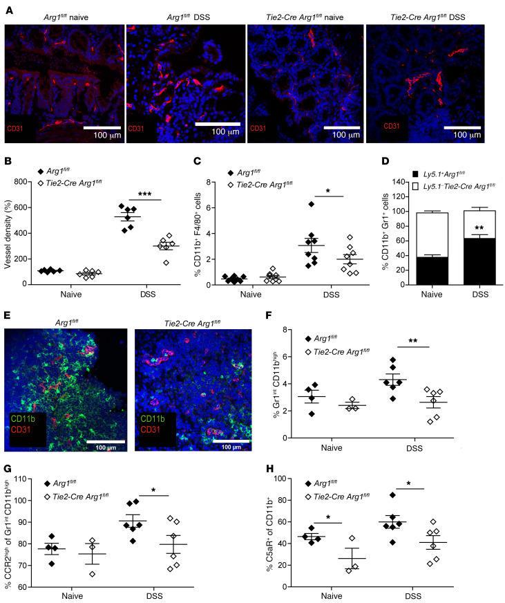
Figure 4. Arg1 increases vessel density and enhances myeloid cell adhesion.
(A and B) Colonic tissue sections of naive or DSS-treated Tie2-Cre Arg1fl/fl and Arg1-expressing littermates (Arg1fl/fl) were stained for CD31 (red) and DAPI (blue) (A), and the relative blood vessel density was quantified in 6 individual mice each (B). Scale bars: 100 μm. (C and D) Distribution of the indicated cell subsets was assessed in the LP by flow cytometry; displayed data represent the results of 8 individually analyzed Tie2-Cre Arg1fl/fl mice and Arg1-expressing littermate controls (Arg1fl/fl) (C) as well as 6 individually assessed mixed Ly5.1+/Ly5.1– Tie2-Cre Arg1fl/fl/WT (Arg1fl/fl) bone marrow chimeric mice (D). (E) Colonic tissue sections of DSS-treated Tie2-Cre Arg1fl/fl and Arg1-expressing littermates (Arg1fl/fl) were stained for CD31 (red) and CD11b (green). Scale bars: 100 μm. (F–H) Surface phenotype of myeloid cells harvested from blood vessels draining intestinal tissues of 3–6 individually analyzed Tie2-Cre Arg1fl/fl mice and respective littermate controls (Arg1fl/fl) was assessed by flow cytometry 12 hours after DSS application (F) along with CCR2 (G) and C5aR expression (H). Data were analyzed using the Mann-Whitney U test. *P ≤ 0.05; **P < 0.01; ***P < 0.001. Error bars indicate SD of the mean.

