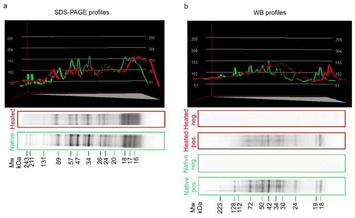
Figure 1.
Colloidal Coomassie-stained 4–20% SDS-PAGE analysis of native and heated (for 60 min at 100 °C) crude (CR) antigen of A. simplex. (a). Western blot (WB) reactivity of anti-A. simplex rabbit IgG antibodies with native and heated (for 60 min at 100 °C) CR antigens of A. simplex. (b). Molecular weight (Mw) estimations in kilodaltons (kDa), densitometric plots of SDS-PAGE and WB profiles were performed using the Bio-1D software (Vilber Lourmat, ver. 15.07, Marne-la-Vallée, France). Green lines in the plots show profiles of the native antigen, while red lines show profiles of the heated antigen. Pos. -strips incubated with hyperimmune serum from a rabbit immunised with native A. simplex CR antigen; Neg. -strips incubated with rabbit pre-immune serum.

