Figure 1.
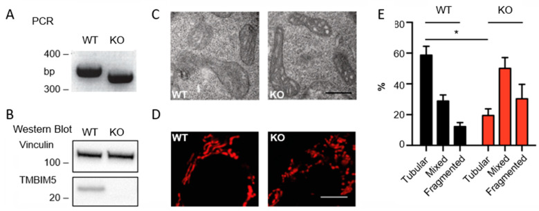
Lack of TMBIM5 results in fragmented and vacuole-like mitochondria. (A) PCR demonstrating a 32 bp deletion in HAP1 TMBIM5-KO (knockout, KO) cells. (B) Immunoblot verifying loss of TMBIM5 protein expression (observed molecular weight at 25 kDa) in HAP1 TMBIM5-KO cells. Vinculin served as loading control, size is indicated. (C) TMBIM5-KO cells exhibit vacuoles, containing low density material and shrunk cristae structures (scale bar: 500 nm) in transmission electron microscopy. (D) Mitochondria in HAP1 wild-type (WT) and TMBIM5-KO cells stained with MitoTracker™ Red CMXRos (scale bar: 3 μm). (E) More than 100 cells from each cell line were imaged by confocal microscopy and categorized according to mitochondrial morphology as tubular, mixed, or fragmented. HAP1 TMBIM5-KO cells have significantly fewer tubular mitochondria, indicated by an asterisk (*). (n = 3 investigators, p < 0.05, graph showing mean ± SD, analyzed with Welch’s t-test).

