Figure 1.
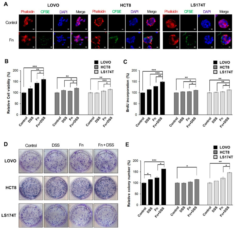
F. nucleatum promotes growth in colorectal cancer (CRC) cells under DSS stimulation. (A) LOVO, HCT8, and LS174T colorectal cancer cells (CRCs) were infected with carboxyfluorescein succinimidyl ester (CFSE)-labeled F. nucleatum (green) for 3 h before analysis using confocal microscopy. The plasma membrane (rhodamine-phalloidin) and nucleus (DAPI) were stained red and blue, respectively. CFSE-labeled F. nucleatum was found mainly in the cytosol or the boundary of the cell; 200 times magnification; scale bar: 10 μm. (B) CRC cells were incubated with the vehicle, DSS, F. nucleatum, or DSS + F. nucleatum. Cell-proliferation rates were evaluated using MTT assays. (C) Proliferation was assessed using a BrdU incorporation assay. Relative changes in the percentage of the absorbance at 450 nm compared to that in the uninfected control. (D) Images showing the effect of F. nucleatum on the colony-forming ability of CRC cells. (E) Graphical representation of the number of colonies formed expressed as percentage of the control. The graph represents relative levels from three independent experiments. The data are presented as the mean ± SEM (* p < 0.05, ** p < 0.01, and *** p < 0.001; unpaired Student’s t-test). Abbreviations: DSS, dextran sodium sulfate; Fn, F. nucleatum.

