FIGURE 3.

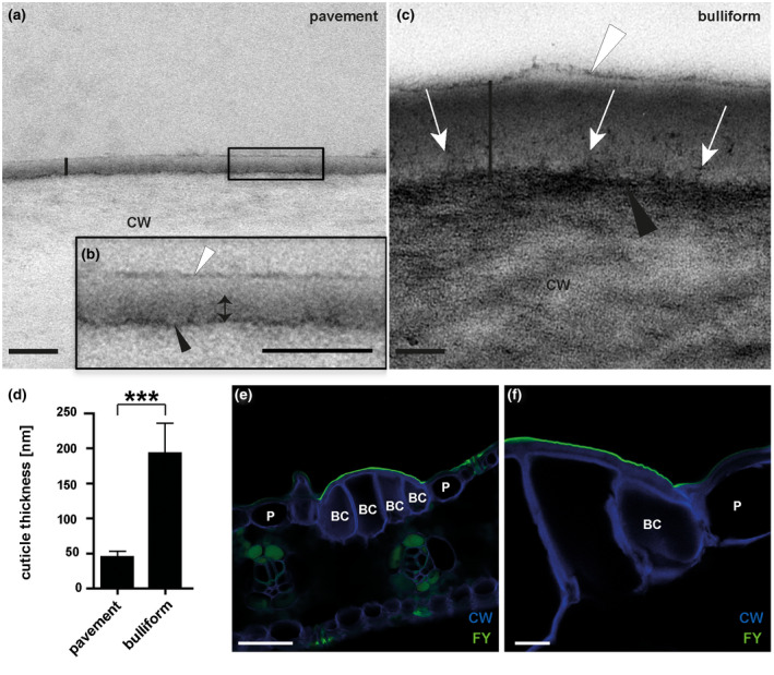
Bulliform and pavement cell cuticles have different thicknesses and ultrastructures. (a‐b) Pavement cell cuticle from a fully expanded adult maize leaf, visualized by TEM (vertical black line marks the full extent of the cuticle), where (b) is the magnified version of the black box in A. Four distinct layers or zones are visible: a thin, darkly stained layer (black arrowhead) at the interface between the cell wall (CW) and cuticle, dark (double headed arrow) and light zones of the cuticle proper, and a darkly stained epicuticular layer (white arrowhead). (c) Bulliform cell cuticle, visualized by TEM (extent marked by vertical black line). The cell wall/cuticle interface (black arrowhead) is diffuse compared to that in pavement cells, and dark‐staining fibrils (white arrows) reach from there into the cuticle. White arrowhead points to the epicuticular layer. Scale bar in (a‐c) = 100 nm. (d) Thickness of different cuticle types, as indicated by the black bars in (a) and (c). Values given as means + SD, n = 45 (three measurements in three different images per cuticle type of five biological replicates). Statistical analysis used two‐tailed unpaired Student's t‐test, with ***p < .001. (e‐f) Fluorol yellow staining of leaf cross‐sections confirms a thicker cuticle over bulliform cells than over the neighboring pavement cells. FY = Fluorol Yellow (lipid stain), CW = Calcofluor White (cell wall counter stain). Scale bar in (e) = 50 nm, in (f) = 10 nm
