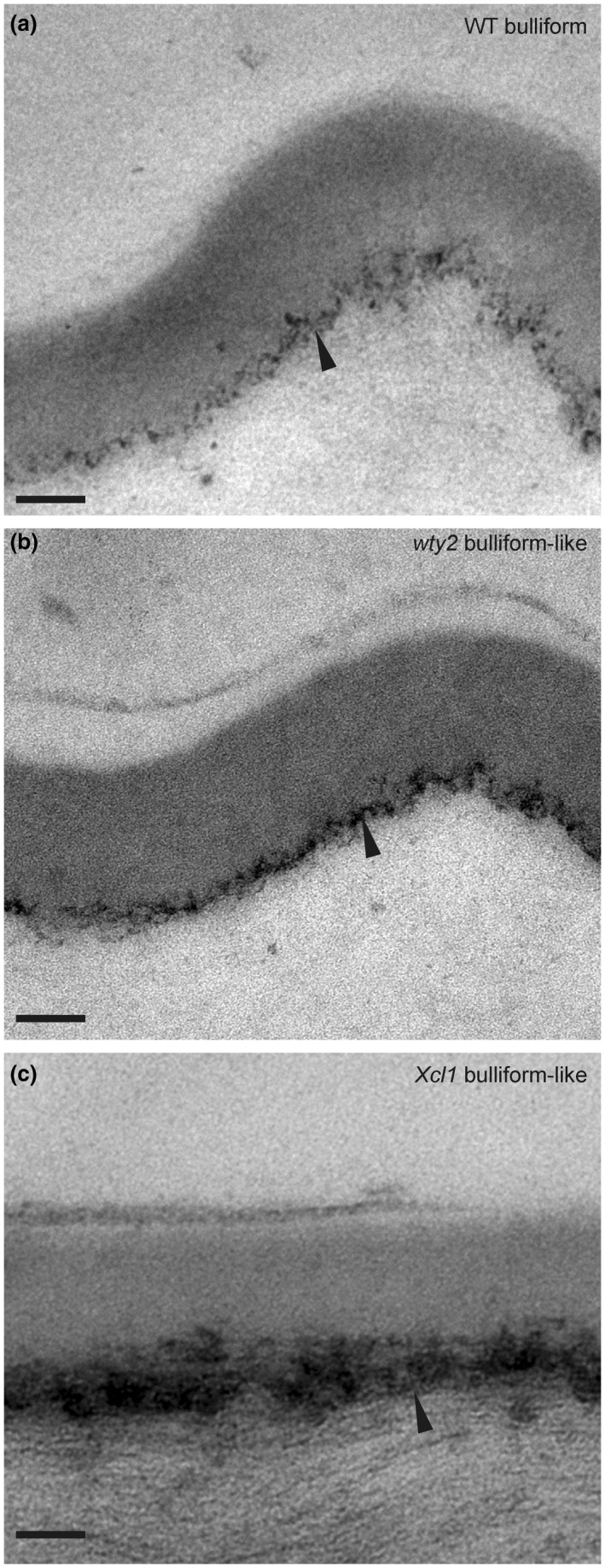
FIGURE 5.

Cuticles of aberrant epidermal cells in bulliform‐enriched mutants display BC‐like ultrastructure. (a‐c) Bulliform cell cuticle of wild‐type (WT), wty2, and Xcl1 mutant, visualized by TEM. Images in (b and c) display the outer surface of abnormal bulliform‐like cells in the respective mutants, identified by the presence of related abnormal epidermal features of the area (warts in wty2, extra cell layer in Xcl1) before acquiring the TEM images. The black arrowhead indicates the diffuse cell wall/cuticle interface of bulliform cells (a) and bulliform‐like cells (b, c). For comparison, images are shown in the same magnification as TEM images in Figure 3a and c. Scale bar = 100 nm
