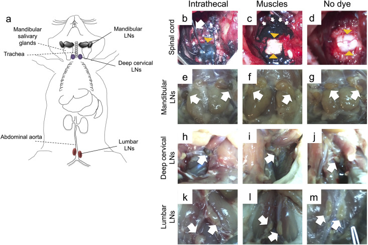
Fig 1. Locating LNs that drain the cervical spinal cord following intrathecal infusion of india ink.
(a) Schematic drawing of rat LNs assessed macroscopically (solid lines) and respective anatomical landmarks (dotted lines). Mandibular LNs, herein referred to as mandibular and accessory mandibular LNs collectively [21], are located adjacent to and rostromedially from the sublingual and mandibular salivary glands. Cranial deep cervical LNs (referred to as deep cervical LNs) are dorsal to the trachea, and lumbar aortic LNs (here referred to as lumbar LNs) are adjacent to the abdominal aorta. (b) India ink was infused intrathecally by a catheter, which was inserted through a small hole in the dura at the C7/T1 level. The white arrow shows the catheter and the yellow arrowhead indicates the spinal cord, which is stained by the dye below the dura. (c) In control rats, the dura remained intact and the dye was allowed to diffuse in the surrounding muscles (white arrows), leaving the spinal cord unstained (yellow arrowheads). (d) In the third group, which received no dye, both the spinal cord (arrowheads) and muscles were unstained. (e-j) (e) Mandibular LNs and (h) deep cervical LNs of rats receiving intrathecal dye were consistently stained blue/black, but these LNs remained unstained in the (f, i) muscle control and the (g, j) no dye groups. (k-m) Lumbar LNs remained unstained in all groups.

