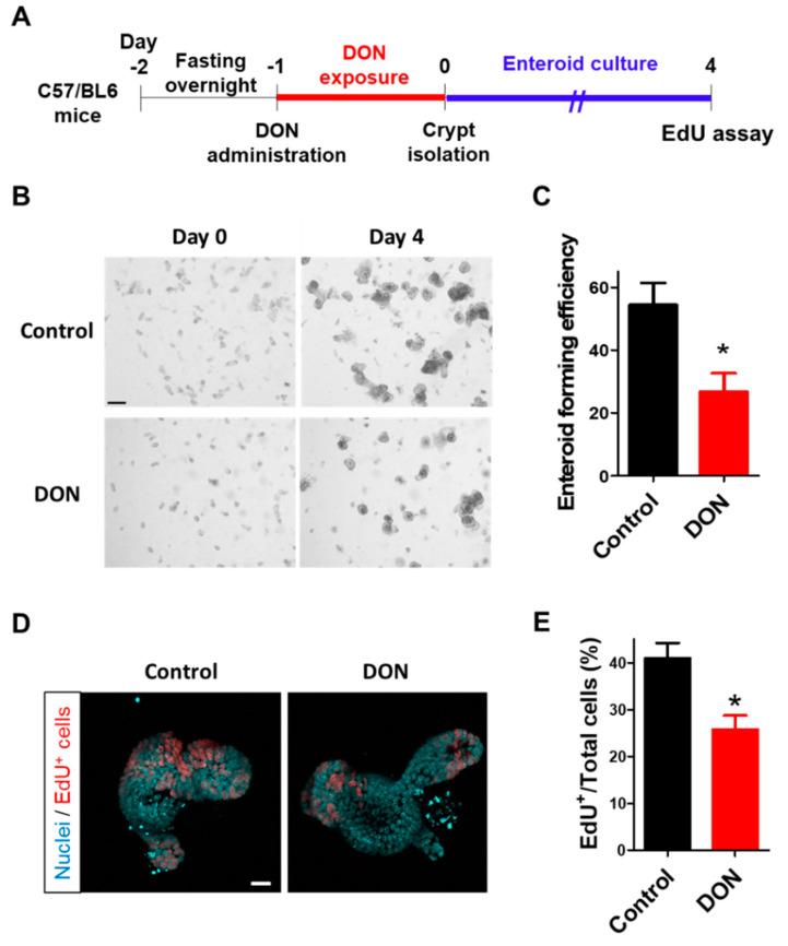
Figure 5.
Effects of oral administration of DON to mice on intestinal stem cells. (A) Scheme of the experimental design. After fasting overnight, Wild-type (WT) mice were orally administered with DON at a dose of 50 mg/kg body weight. After the crypts were isolated from the mice at 24 h after DON exposure, enteroids were prepared and cultured for four days. (B) Representative images of enteroids (at day 0 or 4 after crypt isolation) derived from mice with or without oral DON administration. Scale bar: 200 µm. (C) Enteroid-forming efficiency from mice with or without oral DON administration. Enteroid-forming efficiency was calculated from the ratio of the number of enteroids at day 4 to the number of crypts at day 0. Mean ± SEM, n = 8–12. Asterisk (*) indicates a significant difference. (p < 0.05; Student′s t-test.) (D) Representative confocal images of enteroids at day 4 after crypt isolation. EdU+ cells (red) show proliferative cells and nuclei stained by Hoechst 33342 (sky blue). Scale bar: 20 µm. (E) EdU+ cell quantification in enteroids at day 4 after crypt isolation. The number of EdU+ cells was normalized with the number of total cells and expressed as EdU/Total cells (%). Mean ± SEM, n = 12–18. Asterisk (*) indicates a significant difference (p < 0.05; Students t-test).

