Figure 1.
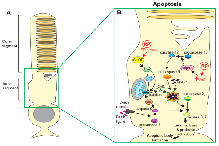
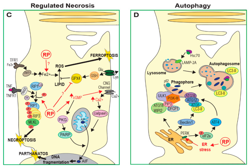
Cell death mechanisms in Retinitis pigmentosa (RP). (A) Schematic of a rod photoreceptor. (B) Both intrinsic pathways (through detection of cell damage) and extrinsic pathways (through interaction with immune cells and death receptors) can lead to apoptosis. Both pathways lead to the release of CytC from mitochondria and oligomerisation of CytC, APAF1 and pro-caspase-9 to form the apoptosome. Activated caspase-9 cleaves executioner caspases 3 and 7. (C) Photoreceptor death can occur by various forms of regulated necrosis. Necroptosis requires activation of RIP1/RIP3, leading to the activation and oligomerisation of MLKL, which forms pores in the cytoplasmic membrane. Parthanatos can occur following activation of PARP, which can be induced by high cGMP levels, leading to nuclear translocation of AIF. Some RP-causing mutations may lead to elevated Fe2+ and/or inhibition of GPX4, causing increased lipid oxidation and death by ferroptosis, although the mechanism is not known. (D) Macroautophagy can be initiated by endoplasmic reticulum (ER) stress via ATG12 activation, direct activation of ATG18/WIP12 and activation of beclin1. Phagophores engulf large cellular components and fuse with lysosomes to form autophagosomes and lysosomal enzymes then degrade the contents of autophagosomes. Proteins also enter lysosomes by hsc-70-assisted chaperone-mediated autophagy via the LAMP1 receptor and by microautophagy through invagination of the lysosomal membrane. (CytC = cytochrome C; APAF1 = apoptotic protease-activating factor 1; RIP = receptor-interacting serine/threonine-protein kinase; MLKL = mixed lineage kinase domain-like pseudokinase; PARP = poly-ADP ribose polymerase; AIF = apoptosis-inducing factor; GPX4 = glutathione peroxidase 4; ATG = autophagy-related protein; LAMP = lysosome-associated membrane glycoprotein).


