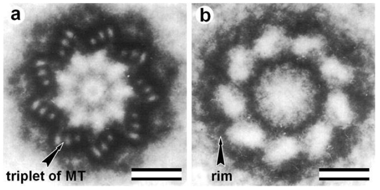
Figure 3.
Rotation images of centriole and centriolar rim. Nine photographs with a rotation angle of 40° were superimposed to obtain images: (a) centriole; and (b) centriolar rim after 1 M KC1 treatment. Scale bar: 100 nm. From [104] with modifications.

